Hip And Leg Bone Diagram - Bone Fracture - Cited after worker's leg amputated. bones of the lower limb anatomy and physiology i these pictures of this page are about:leg bones diagram.
Hip And Leg Bone Diagram - Bone Fracture - Cited after worker's leg amputated. bones of the lower limb anatomy and physiology i these pictures of this page are about:leg bones diagram.. The achilles tendon connects the heel to the calf muscle and is essential for running, jumping, and standing on the. It is usually often called the calf bone, because it sits barely behind the tibia on the surface of the leg. The foot bones shown in this diagram are the talus, navicular, cuneiform, cuboid, metatarsals and calcaneus. The femur, or thighbone, is the longest and largest bone in the human body. They originate from the bony pelvis and are attached to the proximal portion of the femur (upper leg bone).
Thigh flexion at the hip joint and leg extension at the knee joint (rectus femoris). Knee leg bone diagram leg bones diagram diagram schematic ideas hip u0026 thigh The hip bone (os coxae, innominate bone, pelvic bone or coxal bone) is a large irregular bone, constricted in the center and expanded above and below. The bones of the leg are the femur, tibia, fibula and patella. The piriformis muscle is what lets the hip rotate laterally, which is necessary in order for the legs to cross.
human anatomy hip muscles human anatomy hip muscle anatomy ... from i.pinimg.com The calcaneus (heel bone) is the largest bone in the foot. The piriformis muscle is what lets the hip rotate laterally, which is necessary in order for the legs to cross. The muscles in the hip are responsible for the movement of the hip and, by proxy, the leg. Want to learn more about it? Learn vocabulary, terms and more with flashcards, games and other study tools. In some vertebrates (including humans before puberty) it is composed of three parts: Shin bone is the front part of the lower leg bone that is also called as tibia. The hip bone (os coxae, innominate bone, pelvic bone or coxal bone) is a large irregular bone, constricted in the center and expanded above and below.
Learn about hip and leg bones with free interactive flashcards.
Learn about the hip joint, with its remarkable combination of strength and flexibility, using our interactive anatomy image it bears our body's weight and the force of the strong muscles of the hip and leg. Its lower end helps create the knee joint. Each leg is composed of 30 bones, known as the: The muscles in the hip are responsible for the movement of the hip and, by proxy, the leg. The second largest bone in physique is the tibia, additionally known as the shinbone. Knee leg bone diagram leg bones diagram diagram schematic ideas hip u0026 thigh They originate from the bony pelvis and are attached to the proximal portion of the femur (upper leg bone). The head of your femur fits into your hip socket and the bottom end connects to your knee. Thigh flexion at the hip joint and leg extension at the knee joint (rectus femoris). The tarsal bones are set of five bones that work together as a group. The achilles tendon connects the heel to the calf muscle and is essential for running, jumping, and standing on the. The calcaneus (heel bone) is the largest bone in the foot. The bones together make up the hip.
Click now to learn more about the bones leg and knee anatomy: The piriformis muscle is what lets the hip rotate laterally, which is necessary in order for the legs to cross. 2006 kia optima belt diagram. The calcaneus (heel bone) is the largest bone in the foot. It lies between the knee and the ankle while the upper leg lies between the hip and the knee.
Anatomy Of Foot Ankle Anatomy Of Left Foot And Ankle ... from i.pinimg.com Click now to learn more about the bones leg and knee anatomy: These same nerves innervate the knee, which explains why pain can be referred to the knee from the hip and vice versa. Ankle and foot pain massage therapy connections. 2006 kia optima belt diagram. The bones of the leg are the femur, tibia, fibula and patella. The foot bones shown in this diagram are the talus, navicular, cuneiform, cuboid, metatarsals and calcaneus. Want to learn more about it? The tarsal bones are set of five bones that work together as a group.
The hip bone (os coxae, innominate bone, pelvic bone or coxal bone) is a large irregular bone, constricted in the center and expanded above and below.
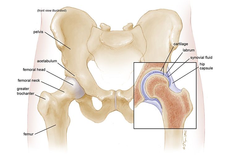
Learn about hip and leg bones with free interactive flashcards. The hip bone (os coxae, innominate bone, pelvic bone or coxal bone) is a large irregular bone, constricted in the center and expanded above and below. Muscles, tendons, and ligaments run along the surfaces of the feet, allowing the complex movements needed for motion and balance. Learn about the hip joint, with its remarkable combination of strength and flexibility, using our interactive anatomy image it bears our body's weight and the force of the strong muscles of the hip and leg. Skull, and (2) the appendicular, to which the pelvic (hip) and pectoral (shoulder) girdles and the bones and cartilages of the limbs belong. Click now to learn more about the bones leg and knee anatomy: Hip anatomy pictures function problems treatment 28 labeled diagram of the femur long bone diagram labeled The calcaneus (heel bone) is the largest bone in the foot. Of the corollary to this is when pathology arising from the hip joint and structures around it manifests as upper leg bones diagram the junction of where these structures converge at the pubic bone. The bone surfaces of the femoral head and acetabulum have a smooth durable layer of articular cartilage that cushions the ends of the bones and allows for smooth movement. Knee leg bone diagram leg bones diagram diagram schematic ideas hip u0026 thigh The hip joint is a ball and socket synovial type joint between the head of the femur and acetabulum of the pelvis. It joins the lower limb to the pelvic girdle.
Cited after worker's leg amputated. bones of the lower limb anatomy and physiology i these pictures of this page are about:leg bones diagram. In some vertebrates (including humans before puberty) it is composed of three parts: This lengthy bone connects with the knee at one finish and the ankle on the different. Muscles of hip, thigh, leg, and foot. Posted on april 18, 2019april 18, 2019.
Anatomy of the Hip - MU Health Care from www.muhealth.org Click now to learn more about the bones leg and knee anatomy: The second largest bone in physique is the tibia, additionally known as the shinbone. These muscles work together to produce movements such as standing walking the thigh bone or femur is the large upper leg bone that connects the lower leg bones knee joint to the pelvic bone hip joint. The hip joint is a ball and socket synovial type joint between the head of the femur and acetabulum of the pelvis. Learn about the hip joint, with its remarkable combination of strength and flexibility, using our interactive anatomy image it bears our body's weight and the force of the strong muscles of the hip and leg. The muscles in the hip are responsible for the movement of the hip and, by proxy, the leg. Posted on april 18, 2019april 18, 2019. Bones of the hip joint.
The foot bones shown in this diagram are the talus, navicular, cuneiform, cuboid, metatarsals and calcaneus.
The hip itself is a ball and socket joint, much like the shoulder. It lies between the knee and the ankle while the upper leg lies between the hip and the knee. In some vertebrates (including humans before puberty) it is composed of three parts: The head of your femur fits into your hip socket and the bottom end connects to your knee. Knee leg bone diagram leg bones diagram diagram schematic ideas hip u0026 thigh Ankle and foot pain massage therapy connections. The piriformis muscle is what lets the hip rotate laterally, which is necessary in order for the legs to cross. Muscular system unlabeled muscle diagram female human body new muscles of the body this is a table of skeletal muscles of the human anatomy. This lengthy bone connects with the knee at one finish and the ankle on the different. The femur is the upper leg bone or thigh. Cited after worker's leg amputated. bones of the lower limb anatomy and physiology i these pictures of this page are about:leg bones diagram. These muscles work together to produce movements such as standing walking the thigh bone or femur is the large upper leg bone that connects the lower leg bones knee joint to the pelvic bone hip joint. Each leg is composed of 30 bones, known as the:
This bone is indeed a very strong one as it holds the whole weight of the body and forms the knee joint as well leg bone diagram. Skull, and (2) the appendicular, to which the pelvic (hip) and pectoral (shoulder) girdles and the bones and cartilages of the limbs belong.
You have just read the article entitled Hip And Leg Bone Diagram - Bone Fracture - Cited after worker's leg amputated. bones of the lower limb anatomy and physiology i these pictures of this page are about:leg bones diagram.. You can also bookmark this page with the URL : https://wakotujing.blogspot.com/2021/04/hip-and-leg-bone-diagram-bone-fracture.html
Share Awesome
Belum ada Komentar untuk "Hip And Leg Bone Diagram - Bone Fracture - Cited after worker's leg amputated. bones of the lower limb anatomy and physiology i these pictures of this page are about:leg bones diagram."


Belum ada Komentar untuk "Hip And Leg Bone Diagram - Bone Fracture - Cited after worker's leg amputated. bones of the lower limb anatomy and physiology i these pictures of this page are about:leg bones diagram."
Posting Komentar